Knee Muscle Anatomy Mri - Atlas of Knee MRI Anatomy - W-Radiology / General anatomy and musculoskeletal system.. Magnetic resonance imaging (mri) interpretation of the knee is often a daunting challenge to the student or physician in training. Click on the links to show each structure. Learn anatomy using a full pacs! The muscles of the knee include the quadriceps, hamstrings, and the muscles of the calf. 4, infrapatellar fat pad of hoffa.
The muscles of the knee include the quadriceps, hamstrings, and the muscles of the calf. Knee muscles need to have both good strength and flexibility. 12 photos of the knee muscle anatomy mri. This mri knee cross sectional anatomy tool is absolutely free to use. Cross sectional anatomy of the knee based on mri :
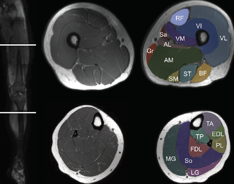
Muscle MRI for Neuromuscular Disorders - Practical Neurology from core4.bmctoday.net Quadriceps tendon semitendinosus tendonsemimembranosus muscle popliteal artery and vein biceps femoris femur vastus medialis sartorius muscle suprapatellar bursa. Scroll through the structures to understand the anatomy. Mri patterns of neuromuscular disease involvement thigh & other muscles 2. Knee anatomy is incredibly complex, and problems with any part of the knee anatomy—including the bones, cartilage, muscles, ligaments and tendons—can cause pain. The main knee muscles are the quadriceps, hamstrings and calf muscles. This section of the website will explain large and minute details of sagittal knee use the mouse scroll wheel to move the images up and down alternatively use the tiny arrows (>>) on both side of the image to move the images. This webpage presents the anatomical structures found on knee mri. Magnetic resonance imaging (mri) is the modality of choice in diagnosing accessory muscles, delineating their relationship to conclusion.
Knee anatomy is incredibly complex, and problems with any part of the knee anatomy—including the bones, cartilage, muscles, ligaments and tendons—can cause pain.
Seems like it should be pretty easy, right? Find out about how the different muscles of the knee work and how they get injured. Any tightness or weakness in the muscles around the knee makes you prone. Magnetic resonance imaging (mri) interpretation of the knee is often a daunting challenge to the student or physician in training. Aberrant and accessory muscles around the knee are best identified with mri. Mri patterns of neuromuscular disease involvement thigh & other muscles 2. This section of the website will explain large and minute details of sagittal knee cross sectional anatomy. This webpage presents the anatomical structures found on knee mri. Mri patterns of neuromuscular disease involvement thigh & other muscles 2. To begin, we use a coronal scan of a left knee. General anatomy and musculoskeletal system. Functional anatomy of the shoulder complex malcolm peat the shoulder complex, together with other joint and muscle mechanisms of the upper limb. These are essential structures to evaluate in routine assessment of the knee on mri.
Abnormal anatomy with normal signal. This mri knee cross sectional anatomy tool is absolutely free to use. Want to learn more about it? The journal of musculoskeletal medicine. By now you probably know that the anatomy is deceptively complex, combinations of injuries can be challenging, and of course the referring clinician's expectations are as high as the range of meniscus injuries is wide.
mri knee anatomy | knee sagittal anatomy | free cross ... from www.mrimaster.com Tips to keep joints healthy. Scroll through the structures to understand the anatomy. Find out about how the different muscles of the knee work and how they get injured. 4, infrapatellar fat pad of hoffa. Tibial tuberosity with distal patella tendon insertion. Learn anatomy using a full pacs! The main knee muscles are the quadriceps, hamstrings and calf muscles. Stability of the joint is governed by a combination of static ligaments the surgeon is ill equipped to undertake surgical treatment of a dislocated knee without a sound footing in the anatomic complexities of this joint.
Scroll through the structures to understand the anatomy.
Anatomy, symptoms, and radiologic evaluation. Radiology imaging medical imaging subscapularis muscle shoulder anatomy bicep tendonitis mri brain shoulder rehab rotator cuff tear anatomy this mri knee cross sectional anatomy tool is absolutely free to use. This webpage presents the anatomical structures found on knee mri. Musculoskeletal radiology south texas radiology group. Articular surface of patella and femur, condyle, epicondyle and muscles (popliteus, sartorius, gastrocnemius, semimembranous with. Learn anatomy using a full pacs! Use the checklist to quiz yourself. Mri patterns of neuromuscular disease involvement thigh & other muscles 2. Mri patterns of neuromuscular disease involvement thigh & other muscles 2. Song, uc san francisco msiv gillian lieberman md. 1 november 2002 mri anatomy of the knee and shoulder james y. Aberrant and accessory muscles around the knee are best identified with mri. Involved early gray = muscle:
By now you probably know that the anatomy is deceptively complex, combinations of injuries can be challenging, and of course the referring clinician's expectations are as high as the range of meniscus injuries is wide. Click now to learn more about the bones, muscles, and soft tissues of these regions at leg and knee anatomy: Mri patterns of neuromuscular disease involvement thigh & other muscles 2. Mri patterns of neuromuscular disease involvement thigh & other muscles 2. Involved early gray = muscle:
Image result for knee joint mri anatomy | Knee mri from i.pinimg.com Knee anatomy is incredibly complex, and problems with any part of the knee anatomy—including the bones, cartilage, muscles, ligaments and tendons—can cause pain. Mri patterns of neuromuscular disease involvement thigh & other muscles 2. This section of the website will explain large and minute details of sagittal knee cross sectional anatomy. This webpage presents the anatomical structures found on knee mri. The main knee muscles are the quadriceps, hamstrings and calf muscles. Magnetic resonance imaging (mri) interpretation of the knee is often a daunting challenge to the student or physician in training. Knee muscles need to have both good strength and flexibility. Free access interactive and dynamic anatomical atlas.
Free access interactive and dynamic anatomical atlas.
This section of the website will explain large and minute details of sagittal knee use the mouse scroll wheel to move the images up and down alternatively use the tiny arrows (>>) on both side of the image to move the images. Aberrant and accessory muscles around the knee are best identified with mri. Articular surface of patella and femur, condyle, epicondyle and muscles (popliteus, sartorius, gastrocnemius, semimembranous with. Anatomy, symptoms, and radiologic evaluation. To begin, we use a coronal scan of a left knee. The muscles of the knee include the quadriceps, hamstrings, and the muscles of the calf. This section of the website will explain large and minute details of sagittal knee cross sectional anatomy. Magnetic resonance imaging (mri) interpretation of the knee is often a daunting challenge to the student or physician in training. The muscles of the knee include the quadriceps, hamstrings, and the muscles of the calf. By now you probably know that the anatomy is deceptively complex, combinations of injuries can be challenging, and of course the referring clinician's expectations are as high as the range of meniscus injuries is wide. Seems like it should be pretty easy, right? Involved early gray = muscle: Knee muscles need to have both good strength and flexibility.


0 Komentar